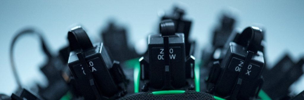
The Sir Peter Mansfield Imaging Centre, University of Nottingham have, in operation, the first 50 channel OPM-MEG array based upon QuSpin sensors. The system uses Gen-2 QZFM mounted on the scalp using a flexible (EEG-like) cap, to measure the magnetic fields generated by current flow through neuronal assemblies. In this way it offers direct and non-invasive inference on human brain electrophysiology. The system is housed within a dedicated magnetically shielded room (MSR) built by Magnetic Shields Limited (MSL). This acts to reduce environmental magnetic interference, as well as limit static (Earths) field. Background fields are further controlled by a set of bi-planar coils (Holmes et al, NeuroImage 2018) – also now available commercially from MSL. This precise field control, coupled with the lightweight scalp mounted sensors and cap, uniquely enables MEG measurement even if a subject moves during the measurement (Boto et al, Nature, 2018).
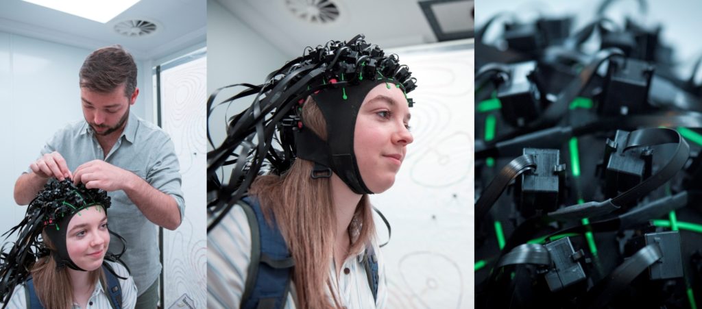
As a first demonstration of our 50 channel device, we employed a visuo-motor task. The subject was shown a visual stimulus comprising a pattern of drifting concentric circles (known as a circular grating). Whilst the visual stimulus was on the screen, the subject was asked to move their finger. Each trial lasted 7 s with the grating on-screen for between 2.5 s and 3 s; in total, 100 trials were presented. Following OPM-MEG data collection, the locations and orientations of the sensors on the scalp were measured using a newly developed optical scanning technique. Accurate knowledge of OPM placement allowed data modelling using a beamformer, to determine precisely where, in the brain, any measurable neuromagnetic effects originate.
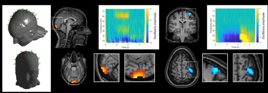
Figure 2 shows the results of these experiments. The left-hand panel shows the locations of the OPMs on the scalp as determined by optical scanning. The centre and right panels show measured brain function in the visual, and motor cortices respectively. We were able to measure high fidelity MEG data which showed that presentation of the visual stimulus elicited an increase in 55-70 Hz “gamma” oscillations in the primary visual cortex. Meanwhile, the finger movement induced a drop in beta oscillations during movement, followed by a post-movement increase above baseline (known as the beta rebound) immediately following stimulation. This beta oscillatory response was shown to be well localised to primary sensorimotor cortex.
These experiments are the first time such a large QuSpin magnetometer array has been operated. Results show that we can achieve high fidelity neuroimaging data with a high sensor count. Combining the sensor array with magnetic shielding and novel coil design means subjects are able to move during data acquisition. Further, the use of optical scanning enables sensor positions to be defined, and consequently accurate localisation of the origins of brain function – in this instance to multiple brain areas in a single experiment. These initial results are a significant step towards a whole head OPM-MEG device. With increased flexibility to adapt to any head shape, movement tolerance to enable novel experimental paradigms, and extremely high-quality data, this offers a step change in neuroscientific experimentation.
The University of Nottingham OPM-MEG group are funded by the Wellcome Trust and the Engineering and Physical Sciences Research Council UK.